Skip to Main Content

Heart Failure

Approximately 6.5 million adult Americans have heart failure. Heart failure, which affects one in five adults age 50 and older over their lifetime.

If you’re experiencing heart failure symptoms (such as difficulty breathing, fatigue or swelling of your legs), time is on your mind. It’s on ours, too. Nearly 4 decades of innovation have resulted in proven therapies for heart failure, which affects one in five adults age 50 and older over their lifetime.

The good news? Early diagnosis, treatment and lifestyle changes can mean a second chance for a longer, more fulfilling life. CHI Health Clinic Heart Institute's comprehensive heart failure program and team of experts offer highly specialized, proven treatments to help patients with heart failure get back to the life they love.

What is Heart Failure?

Heart failure is a chronic, progressive condition that means the heart isn't able to pump enough blood for your body’s needs. This causes fluid retention and congestion of the lungs or other parts of your body such as the ankles, legs and the abdomen. A diagnosis of heart failure does not mean the heart has stopped working. It does mean the heart muscle function has worsened over time.

At first, the heart tries to keep up with its workload by getting larger. The chambers change in shape, size or geometry so they can contract more strongly and pump more blood. The heart may develop more muscle mass because the contracting cells get bigger and the heart is able to pump more strongly, for a while. Blood vessels get narrower to keep blood pressure up. The body diverts blood from less important tissues and organs so it can maintain blood flow to the heart and brain. 

The Heart Failure Management Program at CHI Health provides outpatient treatment and education to help chronic heart failure patients live longer, more rewarding lives. Intravenous therapy, monitoring and education are done in an outpatient setting to avoid hospitalization and decrease overall treatment costs. A multidisciplinary team, consisting of a physician, advanced practice providers, nurses, pharmacists, dietitians and others, participates in the care and support of the patient to obtain optimal therapy outcomes.

Heart Failure Program Highlights

  • Guidelines Directed Medical Therapy Clinic
  • Advanced Heart Failure Clinic
  • Inherited Cardiomyopathies Clinic
  • Amyloid Cardiomyopathy Clinic
  • Inflammatory Cardiomyopathies Clinic
  • Remote Hemodynamic Monitoring Clinic
  • Outpatient Diuretic Infusion Clinic
  • Cardio-Oncology Clinic

Left-Sided Heart Failure

When the heart pumps, it moves oxygen-rich blood from the lungs to the left atrium, and then into the left ventricle, which pumps it throughout the body.

Because the left ventricle provides most of the heart's pumping power, it's larger than the other chambers and vital for normal function. In left-sided or left ventricular (LV) heart failure, the left side of the heart must work harder to pump the same amount of blood.

There are two types of left-sided heart failure.

  • Heart failure with reduced ejection fraction (previously called systolic heart failure): When the heart muscle contracts or beats, it pumps blood out of the heart. In systolic failure, the left ventricle loses its ability to contract normally. The heart can't pump with enough force to push enough blood into circulation.
  • Heart failure with preserved ejection fraction (previously called diastolic heart failure): The heart relaxes between beats, allowing blood to fill up its chambers. In diastolic failure, the left ventricle loses its ability to relax normally and the muscle becomes stiff. The heart can't properly fill with blood during the resting period between each beat.

Right-Sided Heart Failure

The right side of the heart pumps "used" blood that returns to the heart through the veins through the right atrium and into the right lower chamber (ventricle). The right ventricle then pumps the blood back out of the heart into the lungs where it picks up oxygen.

Right-sided or right ventricular (RV) heart failure usually occurs as a result of left-sided failure or high pressures in the pulmonary artery that transfers the blood from the right side of the heart to the lung (pulmonary hypertension). When the left ventricle fails, increased fluid pressure is, in effect, transferred back through the lungs and can damage the heart's right side. When the right side loses pumping power, blood backs up in the body's veins. This usually causes swelling in the legs and ankles (edema).

Congestion in Heart Failure

As the heart's pumping becomes less effective, it works harder. The heart muscle becomes enlarged. The kidneys receive less blood and compensate by filtering less fluid, salt and waste out of circulation and into the urine.

Excess fluid in the blood increases the volume of blood, causing blood pressure to increase. Fluid may build up in the lungs, liver, gastrointestinal tract, and the arms and legs. This is called fluid "congestion" and for this reason doctors call this "congestive heart failure".  As blood pressure increases, it can damage the blood vessels of the heart and kidneys and those throughout the body.

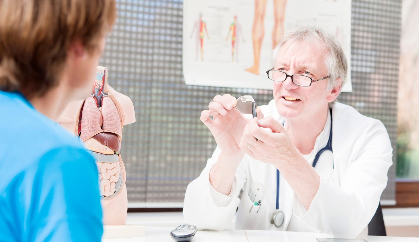
Symptoms of heart failure often begin slowly. Initially, they only occur when you are very active. Over time, you may notice breathing problems and other symptoms even when at rest. Symptoms of heart failure can also occur suddenly, for example after a heart attack or other heart problem. Common symptoms include:

Fatigue

  • Patients with heart failure may feel unusually tired.

Shortness of Breath (Dyspnea)

  • Patients typically report that they run out of breath after exertion. At first, this may begin only when climbing stairs or taking longer walks. Eventually, it can also occur when walking at home. Those who have chest pain or feel that a heavy weight is pressing on their chest should also be screened for possible angina.
  • Orthopnea refers to the shortness of breath that patients can have when lying flat at night. Patients may report having to use a pillow or two under their head and shoulders to sleep. Sitting up with legs hanging over the side of the bed often relieves symptoms.
  • Paroxysmal nocturnal dyspnea (PND) refers to sudden episodes of breathlessness that wake a patient up at night. Symptoms include severe shortness of breath and coughing or wheezing. These usually occur 1-3 hours after falling asleep. Unlike orthopnea, the symptoms may not be relieved by sitting up.

Fluid Retention (Edema) and Weight Gain

  • Patients complain of swelling on the foot, ankle, leg or abdomen. Swelling may occur in the veins of the neck. Fluid retention can lead to sudden weight gain and frequent urination.

Wheezing or Cough

  • Patients may have asthma-like wheezing or a dry, hacking cough that occurs a few hours after lying down but stops after sitting up.

Loss of Muscle Mass

  • Over time, patients may lose muscle weight due to low cardiac output and a significant reduction in physical activity.

Gastrointestinal Symptoms

  • Patients suffer from loss of appetite or a feeling of satiety after eating small amounts. They may also have abdominal pain or bloating.

Pulmonary Edema

When fluid accumulates in the lungs, it is called pulmonary edema. When this happens, the symptoms become worse. These episodes can occur suddenly or gradually build up over the course of days:

  • In addition to shortness of breath, patients sometimes have a cough that produces a pinkish froth.

  • Patients may feel a bubbling sensation in the lungs and feel like they are drowning.

  • Typically, the skin is clammy and pale, sometimes almost blue. This is a life-threatening situation, and the patient should call 9-1-1 or go to the emergency department immediately.

Abnormal Heart Rhythms

  • Patients may have episodes of abnormally fast or slow heart rate.

Central Sleep Apnea

  • This sleep disorder occurs when the brain does not send a signal to the respiratory muscles telling them to breathe. It is common in people with heart failure. Sleep apnea causes disturbed breathing at night. If the heart failure progresses, the apnea can be so acute that a person who cannot breathe wakes up in panic.

What Test for Heart Failure Might I Receive? 

After a physical exam, your doctor may order some tests.

Blood tests are used to:

  • Help diagnose and monitor heart failure
  • Identify risks for heart disease
  • Look for possible causes of heart failure, or problems that may make your heart failure worse
  • Monitor for side effects of medicines you may be taking
  • Check your genes for inherited causes for heart failure.

Echocardiogram

An echocardiogram is often the best test to define the type of heart failure. It is a non-invasive, painless test that uses sound waves to create a moving picture of the heart. The picture is much more detailed than a plain x-ray image and involves no radiation exposure.  

Electrocardiogram

An electrocardiogram (ECG) is a test that measures and records the electrical activity of the heart. It is also called an EKG. An electrocardiogram cannot diagnose heart failure, but it may indicate underlying heart problems. The test is simple and painless to perform. It may be used to diagnose:

  • Previous heart attack
  • Abnormal cardiac rhythms
  • Enlargement of the heart muscle, which may help to determine long-term outlook
  • A finding called a prolonged QT interval may indicate people with heart failure who are at risk for severe complications and therefore need more aggressive therapies.

Myocardial Perfusion Imaging (MPI) Stress Test

The Myocardial Perfusion Imaging (MPI) Stress Test is a noninvasive way to assess the blood flow to the muscle of the heart. We inject a nuclear tracer into the blood stream and the tracer is taken up by the heart muscle cells that receive good blood flow. We image the heart with a camera that senses gamma rays released by the tracer. This basically provides us with a map of where the blood flow to the heart is adequate.

Cardiac MRI

Similar to the echocardiogram, cardiac MRI or CMR is a non-invasive, painless test that uses magnetic resonance to create a moving picture of the heart and provide more comprehensive information about the heart structure.

Cardiopulmonary Exercise Testing

Cardiopulmonary Exercise Testing (CPET) is a metabolic stress test that measures oxygen and carbon dioxide level and assesses cardiorespiratory fitness. It defines and measures the extent of impairment of the heart, lung or peripheral circulation when patients exercise.

Cardiac Catheterization

Cardiac catheterization (sometimes called a coronary angiogram, left heart catheterization or, simply, a “cath”) is an x-ray of the heart while dye fills the coronary arteries. This allows the cardiologist to visualize the coronary arteries to evaluate possible blockage. Right heart catheterization is another type of test that can be done to measure the pressures inside the heart and the amount of blood flow from the heart (cardiac output).

Heart Biopsy

Heart biopsy is also called endomyocardial biopsy or right ventricular biopsy is an invasive testing that is rarely done to identify the cause of heart failure. During endomyocardial biopsy, a small specimen of the heart muscle is obtained and examined under the microscope by a Pathologist to identify the abnormality. 

Heart Failure Treatment

Since heart failure can show in many ways, each individual's treatment may be slightly different. Common treatments for heart failure involve lifestyle changes and medications. Certain procedures may help the heart pump better in some cases of heart failure. Certain procedures such as an angioplasty, cardiac resynchronization devices, valve procedures, or bypass surgeries can help improve heart failure.

If there is a problem with the heart rhythm or conduction, a patient may need a device such as a pacemaker or ICD with or without biventricular pacing (cardiac resynchronization therapy) to help the heart maintain a healthy rhythm and optimize efficiency.

Some procedures are done to treat health problems that may have caused the heart failure such as coronary artery disease. If you have coronary artery disease or valve disease, procedures like a cardiac catheterization or open heart surgery may be done to improve blood flow. This helps the heart pump better, which can improve heart failure symptoms.

For more serious heart failure, other options are available. One option is a left ventricular assist devices (LVAD). LVADs are mechanical devices used to take over the pumping function for one or both of the heart's ventricles, or pumping chambers. A VAD may be necessary when heart failure progresses to the point that medicines and other treatments no longer help. In some cases, a VAD may be used as a bridge to a heart transplant.

A heart transplant replaces the diseased heart with a healthy one from a deceased donor. This is an option for a few people who are very sick. A heart transplant is very serious and not an option for all patients. Your doctor can tell you more.

When treating cancer, some of the treatments may cause damage to your heart, especially if you are at risk of heart disease. Cancer treatments - including chemotherapy drugs and radiation to the chest area - can aggravate existing heart conditions or create new ones. Patients with cancer are more likely to experience long-term heart complications of cancer therapy or die from heart disease. Heart complications that can arise from cancer treatments include heart failure, heart attacks, high or low blood pressures, arrhythmias, and heart valve disease.

The goal of the Cardio-Oncology clinic is to collaborate with your oncologist to make sure you complete the most appropriate cancer therapy, while minimizing possible damage to your heart and treating any heart conditions.

In the Cardio-Oncology clinic, doctors trained in heart disease (cardiologists) will evaluate and treat heart conditions before, during or after cancer therapy, as needed. Patients with a high risk of heart disease, patients with an existing heart disease and patients who develop heart disease or heart complications from cancer treatment will benefit from the clinic services.

What to expect at the Cardio-Oncology clinic

  • Advice and guidance before and during cancer therapy on how to prevent, monitor and treat heart conditions
  • Treatment for pre-existing heart conditions including heart medications if needed
  • Collaborate with oncologist to discuss the cancer treatment options for people at high risk of heart disease or existing heart disease
  • Monitoring heart disease or risk of heart disease before, during and after cancer therapy with regular imaging tests and blood tests to look for signs of heart complications
  • Screening and Treatment of heart conditions that may develop during cancer treatment as a result of certain chemotherapy drugs or receiving radiation therapy to the chest area
  • Lifestyle education to reduce the risk of heart disease including regular exercise, a heart-healthy diet and stopping smoking. 

Are you at risk?


More than 600,000 people die of heart disease every year. Learn about your heart health risk and your heart's real age by taking our online quiz today.

MD Save


Save up to 60% on cardiac imaging with CHI Health through MD Save!

Heart Care Blogs


Read more from our expert providers on heart care topics for you on our CHI Health Better You Blog.